OneNIOS Study Hub
Biology — Class 12 — L15: Circulation of Body Fluids
Lesson-15.pdf). Content covers sections 15.1–15.6.Overview — Why body fluids circulate
Almost all animals have circulating fluids that act as a distributing system (supply substances) and a collecting system (pick up substances) even from the remotest cell. In humans, blood and lymph move nutrients, gases, wastes, hormones and heat, and support defence. This NIOS lesson covers open vs closed circulation, cockroach circulation, human heart and vessels, blood composition and clotting, ABO/Rh and transfusion, blood pressure, lymph, immunity, and disorders (hypertension, atherosclerosis, ECG, pacemaker).
After this lesson you should explain circulatory importance; differentiate open/closed systems; outline cockroach and human organs; describe blood histology and functions; compare artery, vein, capillary; explain clotting, blood groups, BP; describe lymph and immunity types; name immunodeficiency and heart/blood disorders; and state the role of ECG and pacemakers. Notes follow textbook order only. Connect to earlier tissue lessons: blood is fluid connective tissue; cardiac muscle is involuntary striated heart tissue; lymph nodes house lymphocytes already introduced under defence functions of blood.
Section 1: Circulatory system — functions and types (15.1)
Cells need nutrients and oxygen; wastes must leave; hormones travel from glands to targets. Transportation of these substances is circulation. Organs for flow of blood and lymph form the circulatory system.
Functions: (i) nutrients to tissues; (ii) respiratory gases O₂ and CO₂; (iii) metabolic wastes to excretory organs; (iv) hormones to targets; (v) protection by destroying pathogens; (vi) uniform heat distribution.
Open circulatory system: blood does not stay only in closed vessels; flows through parts of the body cavity mixed with body fluid; high pressure not maintained; e.g. prawns, insects (cockroach).
Closed circulatory system: blood in well-defined vessels; high pressure maintained; more efficient; all vertebrates.
| Open | Closed | |
|---|---|---|
| Vessels | Incomplete / sinuses | Continuous tubes |
| Pressure | Low | High |
| Examples | Insects, prawn | Vertebrates |
Section 2: Circulatory system of cockroach (15.2)
Open type: pulsatile heart (dorsal blood vessel) and sinuses. Colourless blood fills the body cavity = haemocoel; blood = haemolymph. Two horizontal septa (dorsal and ventral diaphragms, perforated) divide three sinuses: pericardial (dorsal, encloses heart), perivisceral (viscera), perineural (ventral nerve cord).
Heart: elongated tubular, closed behind, open in front; runs mid-dorsally through thorax and abdomen; thirteen segmentally arranged funnel-shaped chambers; each chamber with a pair of lateral ostia (valved) communicating with pericardial sinus. Anteriorly continues as anterior aorta into head haemocoel. Pair of triangular alary muscles per segment beside the heart. Circulation by contraction/relaxation of heart and alary muscles.
Haemolymph: plasma + haemocytes; no respiratory pigment — does not transport respiratory gases; carries nutrients, maintains hydrostatic pressure, water reservoir.
Section 3: Human circulatory organs (15.3)
Parts: (1) heart — central pump; (2) blood vessels — arteries, veins, capillaries; (3) blood — fluid connective tissue; (4) lymphatic system — nodes and vessels.
3.1 Human heart
Muscular organ of cardiac muscle fibres; fist-sized; four chambers — two upper atria, two lower ventricles (thick walls for long-distance pumping); covered by pericardium.
Valves (open one way; prevent backflow): right AV = tricuspid; left AV = bicuspid (mitral); semilunar valves at origins of aorta and pulmonary artery.
Heart sounds: lubb (1st) — closure of AV valves at atrial/ventricular events; dubb (2nd) — closure of semilunar valves. Cardiac cycle: joint diastole (all chambers relaxed) → atria fill (venae cavae to right atrium; pulmonary veins to left atrium) → atrial systole → ventricular systole → etc.
Conduction system: heartbeat starts at Sino-Atrial (SA) node — modified muscle upper right atrium — the natural pacemaker. Impulse → atria contract → AV node (interatrial septum) → Bundle of His (interventricular septum) → Purkinje fibres in ventricular walls → ventricular systole. Pacemaker influenced by nerves, hormones, CO₂/O₂, heat. Damaged SA node may need artificial pacemaker. ECG (electrocardiogram) records conduction and detects beat disorders.
3.2 Blood vessels
Three wall layers: tunica externa, media, interna. Three kinds:
| Artery | Capillary | Vein | |
|---|---|---|---|
| Direction | Away from heart | Link / exchange | Toward heart |
| Wall | Thick elastic muscular | Single endothelium | Thinner, less muscle |
| Valves | No (except semilunar at heart) | — | Semilunar valves along length |
| Pressure / flow | High, pulsatile, rapid | Falling, slow | Low, non-pulsatile, slow |
| Lumen | Small | Extremely narrow | Large |
| Blood | Usually oxygenated* | Mixed exchange | Usually deoxygenated* |
*Exceptions: pulmonary artery carries deoxygenated blood; pulmonary vein carries oxygenated blood. Arteries → arterioles → capillary beds → venules → veins.
3.3 Path of blood — double circulation
Blood passes through the heart twice in one complete circuit:
- Systemic path: body (deoxygenated) → venae cavae → right atrium → tricuspid → right ventricle → pulmonary arteries → lungs.
- Pulmonary return / systemic output: lungs (oxygenated) → pulmonary veins → left atrium → bicuspid → left ventricle → aorta → body.
Superior vena cava — head and shoulders; inferior vena cava — lower body; both to right atrium. Aorta distributes oxygenated blood from left ventricle.
Intext map: open e.g. insect; closed e.g. human; bicuspid left; tricuspid right; SA node starts beat; capillaries connect arteries to veins; pulmonary vein brings oxygenated blood from lungs; superior vena cava from brain/shoulder region.
Why ventricles have thicker walls than atria: they pump blood much farther — right ventricle to lungs, left ventricle to the whole body — so left ventricular wall is thickest. Pericardium reduces friction as the heart beats in the chest. Lubb–dubb naming: first sound is longer, lower; second is sharper — both from valve closure, not from muscle contraction noise itself. Artificial pacemakers deliver timed electrical pulses when the SA node cannot keep a steady rhythm; ECG is the diagnostic graph that shows P-QRS-T patterns of that electrical journey (textbook level: chamber not contracting properly).
Double circulation keeps oxygenated and deoxygenated streams largely separate in the four-chamber heart — more efficient than the mixed systems of some lower vertebrates. Remember the naming trap: “artery” means away from heart, not “always oxygen-rich”; “vein” means toward heart, not “always oxygen-poor.”
Section 4: Blood — composition, clotting, groups (15.3 continued)
Blood is red, thick, slightly alkaline fluid connective tissue. Importance: transport O₂, nutrients, hormones, wastes to kidney; defence; temperature. Components: plasma ~55% + cellular ~45% (RBC, WBC, platelets). Formation = haemopoiesis in bone marrow.
4.1 Plasma
Pale yellow; water ~90%; proteins albumin, globulin, fibrinogen. Functions: transport digestion products and wastes; hormones; heat distribution; clotting factors (fibrinogen); retain fluid (plasma proteins); acid–base balance; antibodies (immunoglobulins) for immunity.
4.2 Blood cells (Table 15.2 summary)
- Erythrocytes (RBC): ~5,000,000/mm³; marrow; transport O₂ to tissues and much CO₂ to lungs; haemoglobin.
- Leucocytes (WBC): ~4,000–8,000/mm³; granulocytes (neutrophils phagocytosis ~70%; eosinophils; basophils histamine/heparin) and agranulocytes (monocytes phagocytosis; lymphocytes antibodies ~24%).
- Platelets: ~250,000/mm³; fragments without nuclei; initiate clotting.
Disorders: polycythemia (↑RBC); anaemia (↓RBC); leukaemia (↑WBC); leukopenia (↓WBC).
4.3 Coagulation (clotting)
After injury: thromboplastin (from platelets) + prothrombin (plasma) + Ca²⁺ → thrombin; thrombin converts fibrinogen → fibrin (insoluble fibres); fibrin + RBC → clot (scab). Haemophilia — genetic failure of clotting.
4.4 Blood groups and transfusion
Groups A, B, AB, O — genetically fixed for life. Antigens A/B on RBC membrane; antibodies a/b in plasma. Antigen A reacts with antibody b (and B with a) → clumping.
| Group | Antigen on RBC | Antibody in plasma |
|---|---|---|
| A | A | b |
| B | B | a |
| AB | A and B | none |
| O | none | a and b |
Universal donor O (no antigens). Universal recipient AB (no antibodies). Wrong match → agglutination (clumping). Rh factor: Rh⁺ or Rh⁻ protein; Rh⁻ mother with Rh⁺ foetus can produce antibodies risking foetal RBCs if blood mixes.
4.5 Blood pressure
Force of blood on elastic arterial walls. Systolic — ventricular contraction (higher); diastolic — ventricles relaxed/filling (lower). Measured on arm with sphygmomanometer. Healthy adult about 120 ± 5 / 75 ± 5 mm Hg. Difference felt as pulse at wrist; pulse rate ≈ heart rate ~70/min adult.
Plasma as transport medium is more than water: albumin helps hold fluid in vessels; globulins include antibodies; fibrinogen is clotting raw material. Without plasma proteins, fluid leaks more into tissues and clotting fails. RBC count about five million per mm³ carries oxygen; anaemia causes fatigue from poor O₂ delivery. Platelets are fragments, not full cells, but start the cascade at a wound.
Transfusion safety: donor RBCs must not carry antigens that the recipient’s plasma antibodies will attack. That rule explains safe combinations; still learn O to all and all to AB for one-mark questions.
Section 5: Lymphatic system (15.4)
Two circulating fluids: blood and lymph. Continuous exchange between blood capillaries and intercellular fluid; proteins that cannot re-enter blood capillaries return via lymph capillaries to subclavian vein in lower neck. Lymph = modified tissue fluid; clear colourless fluid from capillary walls; contacts body cells. Blister fluid is lymph.
Functions: nutrition/O₂ where blood cannot reach; drain excess tissue fluid; absorb and transport fats from small intestine (lacteals); nitrogenous waste; lymphocytes and antibodies fight bacteria.
| Blood | Lymph |
|---|---|
| Red (haemoglobin) | Colourless |
| Flows rapidly | Very slow |
| RBC, WBC, platelets, plasma | Plasma + WBC mainly |
| Heart → arteries → capillaries → veins → heart | Tissue spaces → lymph capillaries → vessels → subclavian → heart |
System: lymph ducts, nodes, vessels; no pump — muscle movement pushes fluid. Nodes concentrated in neck, armpits, groins; filter bacteria, viruses, cancer cells; house lymphocytes. Spleen (largest lymphoid organ): foetal haemopoiesis; destruction of old RBCs (“graveyard”); blood reservoir; defence. Tonsils also lymphoid.
Section 6: Immunity (15.5)
Immunity — ability to resist harmful disease-producing substances/organisms. Antigen — substance that triggers antibody production (bacteria, viruses, allergens). Antibodies made by lymphocytes.
Natural immunity by birth; acquired during life (exposure or vaccination).
- Active immunity: body makes antibodies after infection or vaccine (weakened germs); long-lasting; e.g. DPT (diphtheria, pertussis, tetanus), BCG (TB); recovery from chickenpox, measles often lifelong.
- Passive immunity: ready-made antibodies injected; short-lived; e.g. anti-tetanus serum (ATS).
Vaccine = small antigen sample too weak to cause disease but enough for antibodies. Vaccines for polio, mumps, measles, tetanus, diphtheria, cholera, etc.
T-cells mature in thymus — identify and destroy antigens directly; lifespan years. B-cells mature in lymphoid tissues (tonsils, spleen) — produce large numbers of antibodies; antibodies short-lived. Lack of T, B or both → high infection risk.
Immunodeficiency: SCID — absence of both T and B from birth; AIDS — HIV destroys T-cells/immune system. HIV routes: sexual contact; contaminated blood; shared needles; mother to foetus via placenta.
Immunity vocabulary for short answers: antigen triggers response; antibody is the protein product; vaccine is controlled antigen exposure; natural immunity is by birth while acquired builds during life. T-cells identify and destroy; B-cells manufacture antibodies. SCID is combined defect from birth; AIDS is acquired through HIV. Active means you make antibodies (often lifelong); passive means you receive ready-made antibodies (temporary) — the usual mark-scheme contrast.
Section 7: Disorders of blood and heart (15.6)
Hypertension — BP above normal (~120/75); linked to stress, overweight, age, diet. Atherosclerosis — fat deposits (atheroma) on arterial inner wall; narrows lumen, impairs heart blood supply. Arteriosclerosis — hardening/loss of flexibility of arterial walls with age and deposits. Treatments: balloon angioplasty with stent; heart bypass with grafted vessel.
ECG records heartbeat as a graph so doctors see which chamber contracts/relaxes wrongly. Pacemaker devices regularise abnormal rhythm when SA node fails.
Distinguish atherosclerosis (fatty plaque / atheroma narrowing lumen) from arteriosclerosis (wall hardening and stiffness). Both impair coronary flow; both appear in terminal “name two heart-related disorders.” Hypertension is pressure too high, not the same as plaque, though they often coexist in real patients. Balloon angioplasty widens the lumen with a stent; bypass grafts a new path around a blocked segment — plumbing metaphors help memory without needing surgical detail.
Section 8: Exam checklist and walkthrough
Open vs closed; cockroach haemocoel/haemolymph/no gas pigment; human 4 chambers, valves, SA→AV→His→Purkinje, lubb–dubb; double circulation path; artery/vein/capillary table; plasma proteins; RBC/WBC/platelets; clotting cascade; O universal donor, AB recipient; Rh pregnancy risk; BP 120/75; lymph vs blood; spleen functions; active vs passive immunity; SCID/AIDS; hypertension/atherosclerosis/arteriosclerosis; ECG/pacemaker.
Terminal-style: functions of RBC, platelets, plasma; clotting flowchart; why AB is universal recipient; systolic vs diastolic + normal values; three lymph vs blood differences; immunity active vs passive; define hypertension and atherosclerosis; what ECG does.
One-line keys: haemopoiesis; albumin/globulin/fibrinogen; platelets clot; transfusion; antigens on RBC antibodies in plasma; group O receives only O; sphygmomanometer; lymph colourless from capillaries; lymph nodes + lymphocytes; spleen/tonsils; SCID and AIDS; T and B cells; hypertension and atherosclerosis.
Integrated story: closed high-pressure circuit pumps oxygenated blood from left heart via aorta to capillaries; plasma filters to tissue fluid; lymph returns excess and fats; right heart sends deoxygenated blood to lungs; clotting seals wounds; ABO/Rh matching prevents agglutination; immune cells and antibodies defend; modern medicine uses ECG, pacemakers, angioplasty when the pump or vessels fail.
Use Formula Sheet lock boxes; drill 10 MCQs and 20 flashcards. Prioritise double-circulation path, vessel comparison, ABO table, clotting steps, and active/passive immunity.
Section 9: Extra depth for 3000-word coverage
Why closed systems are “more efficient”: blood stays in vessels at higher pressure, so delivery to distant tissues is faster and more controlled than bathing organs in a slow haemocoel stream. Cockroach trades respiratory gas transport (done by tracheal system, not blood) for a simpler open design — exam answer should mention both haemolymph functions and lack of respiratory pigment.
Cardiac cycle timing: atria fill during joint diastole; atrial systole tops up ventricles; ventricular systole ejects blood; valves ensure one-way flow — like doors that only open downstream. Chordae tendineae and papillary muscles (seen in internal figures) prevent AV valves from inverting under pressure.
Capillary beds are the business end of the system: only there can exchange of O₂, CO₂, nutrients and wastes occur by diffusion across a single cell layer. Arteries and veins are highways; capillaries are local streets. High total capillary volume explains slow flow and long time for exchange.
Clotting must stay local: cascade amplification is powerful but regulated so free-flowing blood does not solidify. Haemophilia shows how missing one factor collapses the cascade. Calcium ions are cofactors — low Ca²⁺ impairs clotting in theory questions.
Transfusion logic: never give a recipient an antigen their antibodies will attack. Group O cells have no A/B antigens, so any plasma type is safer with O cells (universal donor). Group AB plasma has no anti-A/anti-B, so AB can accept any cell antigens (universal recipient). Always still match Rh in practice.
Lymph nodes as filters: immune surveillance stations where lymphocytes meet pathogens. Swollen nodes in infection are working hardware, not just “sickness signs.” Spleen dual role as blood reservoir and RBC quality control (graveyard) appears often in short answers. Lymph moves slowly without a heart pump; skeletal muscle activity and one-way valves keep it progressing toward the veins of the neck. That is why prolonged immobility can contribute to swelling from fluid pooling — a practical link to tissue fluid balance.
Active immunity is memory: after vaccine or disease, specific clones persist. Passive immunity is rental antibodies — useful in emergency tetanus risk but fades. HIV targets helper T-cells, collapsing both cell-mediated and antibody arms — hence “syndrome” of many infections (AIDS).
Lifestyle and vessels: chronic high fat diet → atheroma → narrowed coronary arteries → angina or heart attack risk; hypertension damages vessel walls. Balloon angioplasty and bypass are engineering fixes for plumbing problems the textbook names clearly for awareness, not surgical detail.
Closed-book drill: (1) six circulatory functions; (2) open vs closed with examples; (3) cockroach heart and haemolymph role; (4) four heart valves and locations; (5) conduction path; (6) double circulation flowchart; (7) artery/vein/capillary table; (8) plasma and cell components; (9) clotting steps; (10) ABO + universal donor/recipient; (11) BP values and instrument; (12) lymph functions; (13) active vs passive immunity; (14) SCID vs AIDS; (15) three vascular/heart disorders + ECG. Completing these covers Lesson 15 terminal exercises.
Quick formula strip: open vs closed; SA→AV→His→Purkinje; body→RA→RV→lungs→LA→LV→aorta; artery away/vein toward; plasma 55%; clot = fibrin mesh; O donor / AB recipient; BP 120/75; lymph return; active vs passive; HIV→AIDS; atheroma fat deposit. Say each with one diagram or table location. This chapter is heavy on definitions and pathways — flashcards repay time better than rereading alone. Also lock: haemopoiesis in marrow; polycythemia/anaemia/leukaemia/leukopenia name pairs; haemophilia genetic clotting failure; pulse rate equals heart rate about seventy per minute in a resting adult. Superior vena cava drains head and shoulders; inferior vena cava drains the lower body; both empty into the right atrium before blood is sent to the lungs for gas exchange and then returned for systemic distribution through the aorta. Review the cockroach section once more: colourless haemolymph, thirteen-chamber heart, ostia, alary muscles, and three sinuses of the haemocoel — a favourite short-answer cluster separate from the human path. You are exam-ready when that insect list and the human double-circulation flowchart both come without notes on the day of the full NIOS senior secondary biology examination paper for this academic year period.
MCQ Quiz — L15 Circulation of Body Fluids
Flashcards — L15
Golden Rules — L15 Circulation of Body Fluids
Most exam-important points from this chapter:
Open vs closed & cockroach
Open: sinuses, low pressure (insect haemocoel). Closed: vessels, high pressure (us). Cockroach heart 13 chambers, ostia, alary muscles; haemolymph does not carry O₂.
Heart pump & path
SA node → AV → His → Purkinje. Tricuspid right, bicuspid left. Double circulation: body→R heart→lungs→L heart→body. Pulmonary artery deox; pulmonary vein ox.
Vessels & blood
Artery away thick high P; vein toward valves low P; capillary exchange. Plasma 55%; RBC O₂; WBC defence; platelets clot. Cascade: prothrombin→thrombin→fibrin.
Groups & BP
O universal donor; AB universal recipient. Match antigens/antibodies. Rh matter in pregnancy. BP ~120/75; pulse ≈ heart rate.
Lymph, immunity, disease
Lymph drains tissue fluid & fats; nodes filter; spleen graveyard. Active vs passive immunity; T and B cells. SCID/AIDS; hypertension/atherosclerosis; ECG & pacemakers.
Past Year Questions — L15 Circulation of Body Fluids
PE-only questions for this chapter only. 3 item(s). No overlap with other lessons. Tap Show answer after you try each question.
Q1. Heart is three-chambered in reptiles. But a partially divided ventricle (i.e., four-chambered heart) is present in
Why it clicks: Crocodiles have a four-chambered heart; most other reptiles are three-chambered.
Q2. (a) Write the differences between blood and lymph. (b) With the help of flowchart, only show the route of blood flow and lymph flow in the human body. 3 (H) a
This question needs a diagram — open the answer to view the HD model figure.
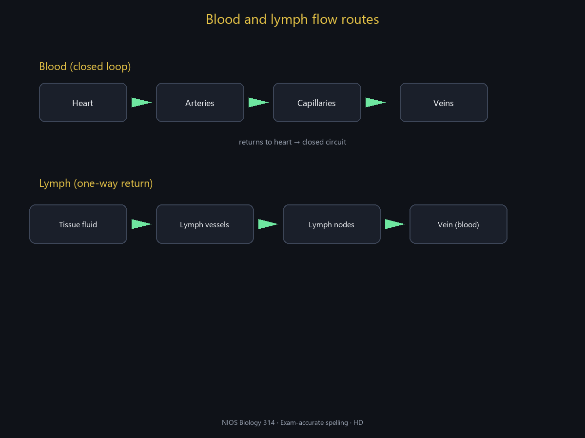
Why it clicks: Lymph is one-way return of fluid the blood leaked; blood is a closed loop.
Q3. The blood protein that provides factors for clotting is
Why it clicks: Plasma protein converted to fibrin mesh in a clot. Thrombin is the enzyme, not the main clot protein store.
Problem Solving — L15 Circulation of Body Fluids
Six problems spanning this chapter’s NIOS Biology (314) syllabus. Every question is built from the notes and formula sheet: solve with definitions and equations first, then read the formal textbook-style write-up, the easy explanation, and the topic in depth (key relations, meaning, exam tips). If the question says draw, a labelled pencil sketch is provided. Explanations open by default.
List the three formed elements of blood and one function of each.
Pencil sketch (labelled)
Solution — step by step
- Erythrocytes (RBC) — transport O₂ (haemoglobin).
- Leucocytes (WBC) — immunity/defence.
- Platelets (thrombocytes) — clotting.
Final answer: RBC O₂; WBC defence; platelets clot
Key relations / definitions
Textbook formal language
Blood is a fluid connective tissue with plasma matrix and cellular elements.
Key relations: Plasma + formed elements; RBC O₂; WBC defence; platelets clotting. State the definition or law first (NIOS style), use correct biological terms, and end with a clear boxed conclusion.
Easy language (same idea, plain words)
Red cells carry oxygen, white cells fight germs, platelets plug bleeds.
Read the question once for the idea, once for the details. Write the definition or equation, then apply it. Check labels and units if any numbers appear.
Topic in depth — Composition of blood
Plasma carries nutrients, hormones, CO₂ as bicarbonate, proteins.
Linked to chapter notes (L15). Remember: Plasma + formed elements; RBC O₂; WBC defence; platelets clotting. Most exam errors mix up similar terms, reverse cause and effect, or skip labelled diagrams.
Exam tip
Open with a one-line definition, then use: Plasma + formed elements; RBC O₂; WBC defence; platelets clotting. For diagram questions, label every part asked and keep lines neat.
Common mistakes
- Confusing prokaryote with eukaryote (or plant with animal tissues).
- Mixing up similar pathways (e.g. photosynthesis vs respiration; mitosis vs meiosis).
- Writing vague answers without key technical terms from NIOS notes.
- Forgetting to label diagrams or state units where numbers are used.
What is double circulation in humans? Why is it advantageous?
Pencil sketch (labelled)
Solution — step by step
- Blood passes twice through the heart per full body circuit: pulmonary and systemic.
- Keeps oxygenated and deoxygenated blood largely separate; efficient O₂ delivery to tissues.
Final answer: Two circuits via heart; efficient O₂ supply
Key relations / definitions
Textbook formal language
Four-chambered heart enables complete double circulation in mammals/birds.
Key relations: Pulmonary + systemic circuits; Right heart → lungs; left heart → body. State the definition or law first (NIOS style), use correct biological terms, and end with a clear boxed conclusion.
Easy language (same idea, plain words)
Heart pumps to lungs, gets O₂, returns, then pumps to body—two loops.
Read the question once for the idea, once for the details. Write the definition or equation, then apply it. Check labels and units if any numbers appear.
Topic in depth — Double circulation
Separate oxygenated and deoxygenated streams raise efficiency of delivery.
Linked to chapter notes (L15). Remember: Pulmonary + systemic circuits; Right heart → lungs; left heart → body. Most exam errors mix up similar terms, reverse cause and effect, or skip labelled diagrams.
Exam tip
Open with a one-line definition, then use: Pulmonary + systemic circuits; Right heart → lungs; left heart → body. For diagram questions, label every part asked and keep lines neat.
Common mistakes
- Confusing prokaryote with eukaryote (or plant with animal tissues).
- Mixing up similar pathways (e.g. photosynthesis vs respiration; mitosis vs meiosis).
- Writing vague answers without key technical terms from NIOS notes.
- Forgetting to label diagrams or state units where numbers are used.
Define systole and diastole. Which valves close to produce the first heart sound (lub)?
Pencil sketch (labelled)
Solution — step by step
- Systole: chamber contraction; diastole: relaxation/filling.
- First sound (lub): closure of atrioventricular valves (tricuspid & bicuspid/mitral).
Final answer: Contraction/relaxation; AV valves → lub
Key relations / definitions
Textbook formal language
Cardiac cycle is coordinated contraction sequence ensuring one-way flow.
Key relations: Systole = contraction; Diastole = relaxation; Lub-dub valves. State the definition or law first (NIOS style), use correct biological terms, and end with a clear boxed conclusion.
Easy language (same idea, plain words)
Squeeze is systole, rest/fill is diastole; lub is AV valves slamming shut.
Read the question once for the idea, once for the details. Write the definition or equation, then apply it. Check labels and units if any numbers appear.
Topic in depth — Systole and diastole
Dub is semilunar valves (aortic/pulmonary) closing.
Linked to chapter notes (L15). Remember: Systole = contraction; Diastole = relaxation; Lub-dub valves. Most exam errors mix up similar terms, reverse cause and effect, or skip labelled diagrams.
Exam tip
Open with a one-line definition, then use: Systole = contraction; Diastole = relaxation; Lub-dub valves. For diagram questions, label every part asked and keep lines neat.
Common mistakes
- Confusing prokaryote with eukaryote (or plant with animal tissues).
- Mixing up similar pathways (e.g. photosynthesis vs respiration; mitosis vs meiosis).
- Writing vague answers without key technical terms from NIOS notes.
- Forgetting to label diagrams or state units where numbers are used.
Give three differences between artery and vein.
Solution — step by step
- Arteries carry blood away from heart; veins toward heart.
- Arteries thicker muscular/elastic walls; higher pressure.
- Veins have valves; arteries generally do not (except base of aorta/pulmonary).
Final answer: Away vs toward; thick wall vs valves; pressure high vs low
Key relations / definitions
Textbook formal language
Vessel structure matches pressure and direction of flow.
Key relations: Artery: thick elastic, away from heart; Vein: valves, toward heart; Capillary: exchange. State the definition or law first (NIOS style), use correct biological terms, and end with a clear boxed conclusion.
Easy language (same idea, plain words)
Arteries are high-pressure hoses from the heart; veins are return pipes with doors (valves).
Read the question once for the idea, once for the details. Write the definition or equation, then apply it. Check labels and units if any numbers appear.
Topic in depth — Artery vs vein
Pulmonary artery carries deoxygenated blood—exception to “artery = oxygenated”.
Linked to chapter notes (L15). Remember: Artery: thick elastic, away from heart; Vein: valves, toward heart; Capillary: exchange. Most exam errors mix up similar terms, reverse cause and effect, or skip labelled diagrams.
Exam tip
Open with a one-line definition, then use: Artery: thick elastic, away from heart; Vein: valves, toward heart; Capillary: exchange. For diagram questions, label every part asked and keep lines neat.
Common mistakes
- Confusing prokaryote with eukaryote (or plant with animal tissues).
- Mixing up similar pathways (e.g. photosynthesis vs respiration; mitosis vs meiosis).
- Writing vague answers without key technical terms from NIOS notes.
- Forgetting to label diagrams or state units where numbers are used.
State two functions of lymph.
Solution — step by step
- Returns excess tissue fluid and proteins to blood.
- Transports fats (lacteals) and participates in immune defence (lymph nodes).
Final answer: Fluid return + immunity/fat transport
Key relations / definitions
Textbook formal language
Open lymphatic drainage complements closed blood circulation.
Key relations: Lymph = tissue fluid in lymph vessels; Returns proteins/fluid; immunity. State the definition or law first (NIOS style), use correct biological terms, and end with a clear boxed conclusion.
Easy language (same idea, plain words)
Lymph is the “overflow drain” and immune checkpoint fluid.
Read the question once for the idea, once for the details. Write the definition or equation, then apply it. Check labels and units if any numbers appear.
Topic in depth — Lymphatic system
Lymph is not the same as blood—lacks RBCs.
Linked to chapter notes (L15). Remember: Lymph = tissue fluid in lymph vessels; Returns proteins/fluid; immunity. Most exam errors mix up similar terms, reverse cause and effect, or skip labelled diagrams.
Exam tip
Open with a one-line definition, then use: Lymph = tissue fluid in lymph vessels; Returns proteins/fluid; immunity. For diagram questions, label every part asked and keep lines neat.
Common mistakes
- Confusing prokaryote with eukaryote (or plant with animal tissues).
- Mixing up similar pathways (e.g. photosynthesis vs respiration; mitosis vs meiosis).
- Writing vague answers without key technical terms from NIOS notes.
- Forgetting to label diagrams or state units where numbers are used.
Why is blood group O often called a universal donor for RBCs (simplified school level)?
Pencil sketch (labelled)
Solution — step by step
- Group O RBCs lack A and B antigens.
- Less likely to be agglutinated by recipient anti-A/anti-B (classic textbook simplification).
Final answer: O RBCs lack A/B antigens
Key relations / definitions
Textbook formal language
Transfusion compatibility depends on antigen–antibody matching; Rh also critical.
Key relations: A,B antigens; anti-A/B antibodies; Rh+ has D antigen. State the definition or law first (NIOS style), use correct biological terms, and end with a clear boxed conclusion.
Easy language (same idea, plain words)
O red cells don’t wear A/B flags, so many receivers don’t attack them (school model).
Read the question once for the idea, once for the details. Write the definition or equation, then apply it. Check labels and units if any numbers appear.
Topic in depth — ABO and Rh
Modern practice matches carefully; AB is classic universal plasma/recipient story—don’t mix up.
Linked to chapter notes (L15). Remember: A,B antigens; anti-A/B antibodies; Rh+ has D antigen. Most exam errors mix up similar terms, reverse cause and effect, or skip labelled diagrams.
Exam tip
Open with a one-line definition, then use: A,B antigens; anti-A/B antibodies; Rh+ has D antigen. For diagram questions, label every part asked and keep lines neat.
Common mistakes
- Confusing prokaryote with eukaryote (or plant with animal tissues).
- Mixing up similar pathways (e.g. photosynthesis vs respiration; mitosis vs meiosis).
- Writing vague answers without key technical terms from NIOS notes.
- Forgetting to label diagrams or state units where numbers are used.